Teledermatology Documents
Over the past three years, I have attempted to document as many of the ideas and accomplishments related to the VISN 2 Teledermatology Project as possible before they get lost to memory. Some of this material, such as the manuals used to train the staff to acquire diagnostic images and a description of the Distributed Specialty Care Model, is in presentable form and is included here. Other material is in need of more work and will be made available as time allows.
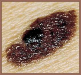
Unlike other medical applications where special cameras capture images under well-controlled conditions, dermatologic imaging has to contend with COTS devices, hand-held operation, and appreciable variations in room illumination. Nonetheless, experience has shown that even these cameras, in concert with a moderate amount of training, can produce images that possess level of quality required for remote diagnosis. The camera that was selected initially for the teledermatology project was the Canon Digital Rebel 300D. This camera was chosen because most of its imaging parameters could be manually set, it came with an excellent macro flash unit, and for the time, it had a fairly high pixel resolution (plus, one was available in the clinic).
The combination of a detailed manual and one-on-one hands-on training resulted in excellent compliance with the imaging protocols and produced diagnostic-quality results across a wide range of caregiver (in)experience. At 26 pages, the manual is not a quick read but it has served as a reference during training and a resource during emergencies. The manual provides each user with an overview and background that affords a comfort with the unit as well as promotes at least a cursory familiarity with all of the camera settings. Photographic performance is improved because the user obtains an understanding not commonly available from the manufacturer’s terse documentation. In addition, presenting a rationale for the selection of each of the camera settings integrates the use of the camera with the clinical protocols in a manner that provides meaning and promotes retention. For example, the training includes an explanation of the benefits of twin macro flash lighting for improved image quality. Detailed control of the macro flash unit and manual access to the camera settings enables the illumination to be set at a high enough level to eliminate most variation (chromatic contamination) due to room lighting while, at the same time, allows the aperture to be stopped down and thereby increase the depth of field (reducing defocus blur) and for the shutter speed to be increased which reduces motion blur. With control over the flux levels, the usual sensitivity reciprocities cease to be an issue in diagnostic image capture. As experience was gained in the clinic, the manuals and the protocols were altered in a continuous adjustment process to meet user capabilities and clinical need.
The writing and proofing of each camera manual required a considerable effort. As indicated above, it was a continuous process of adaptation and revision. The process was also ongoing for another reason: consumer cameras have a very short market life. Consider the changes over a seven year period in one of the most popular digital camera lines, the Canon Digital Rebel series (and their market life): 300D (18 mo.), 350D (18 mo.), 400D (17 mo.), 450D (14 mo.), 500D (11 mo.), and most recently the 550D. Over this period there were more changes in these cameras that needed to be accommodated than just the 6 to 18 Mpixel increase in resolution. Consider the manuals for the first camera used in the clinics (300D) and its successor (350D). Peruse the parameter setting descriptions and note how much changed in the camera controls between two successive iterations of units in the same product line. This is the tip of the iceberg when considering all the modifications in the electronic processing that constitute such a large part of consumer digital cameras today, much of which has no public documentation and is beyond the control of the user.
In addition to providing a manual that integrates camera settings and clinical protocols, there are other utilities that can assist performance in the clinic. Helpful as the paper manuals are, they do not provide the format that is most useful when there is ‘something wrong’ with the camera and there is a patient in the exam room and a dozen more in chairs. Frequently, just picking up a camera can activate any of the score of buttons that coat its surface. What is needed is an online utility that captures the configuration and settings of the camera and the content of its displays. So when the camera won’t take a picture of sufficient quality, calling up a diagnostic utility can provide an intuitive troubleshooting process. With the camera locked up and a less than meaningful right-pointing finger flashing on its LCD, it would be more efficient to be able to click on an equivalent icon on the diagnostic utility’s display on the computer than to start leafing through a manual. Besides coming to the rescue, such a utility could also serve as a camera initialization control. The ability to confirm the initial camera configuration at the start of use can help avoid irretrievable data loss due to incorrect settings that are found out only after the patient has left the clinic. Another utility that can provide a critical service is one that provides image quality assessment of the patient images before the patients leave the exam room. This capability would reduce additional call backs when, on occasion, the standard settings are not adequate or appropriate to capture the necessary diagnostic information. Besides these GUI prototypes, aids to reduce the effects of hand-held operation have been shown to be desirable. With new imaging devices, techniques and staff, there will always be a need for improved workflow assistance.
While the capture of diagnostic quality images with ease and regularity is a core competency of teledermatology, there is much more that goes into making an efficient and effective system. A series of technical reports is being assembled based on the teledermatology experiences at the VA. A more detailed description of the Distributed Specialty Care model will be presented along with those of a number of allied support services. The reports will summarize how all the levels of care come together in a system that provides review and feedback, making teledermatology an interactive, ongoing learning experience. Examples also will be presented that demonstrate how administrative and regulatory problems can be damaging to teledermatology’s prospects. A brief outline of the Distributed Specialty Care model is also available that describes the mechanisms that enable the empowerment of non-specialty clinical staff to expand the accessibility of dermatologic care.