mhtml:file://C:\Documents%20and%20Settings\brian\Desktop\brianwebsite\docs\LiveMicroImagingwithOptiScanStratum.mht!http://www.lawrenz.com/images/nav_linksoben_0.gif

mhtml:file://C:\Documents%20and%20Settings\brian\Desktop\brianwebsite\docs\LiveMicroImagingwithOptiScanStratum.mht!http://www.lawrenz.com/images/schrift.gif
OptiScan Stratum

Welcome!

Bibliography

Info Order

Free Download

Events

Links

Lawrenz Medizinelektronik In Assocation With amazon.com

mhtml:file://C:\Documents%20and%20Settings\brian\Desktop\brianwebsite\docs\LiveMicroImagingwithOptiScanStratum.mht!http://www.lawrenz.com/images/nav_space_0.gif

mhtml:file://C:\Documents%20and%20Settings\brian\Desktop\brianwebsite\docs\LiveMicroImagingwithOptiScanStratum.mht!http://fastcounter.linkexchange.com/fastcounter?187516+375039

Live Micro Imaging with OptiScan Stratum

Optiscan Stratum Breaking new ground in the use of Live Micro Imaging (LMI) technology for skin examination.

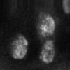
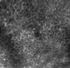
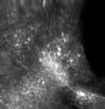
Designed to sit comfortably in the hand, the tip of the Stratum scanner is simply placed against the skin on any part of the body. It uses visible and/or near infrared light to non-invasively produce high magnification (~1000x) images of microscopic cellular structures. Clear subsurface images can be isolated from within living tissue, layer by layer. The digital images of cells appear in real time on the computer screen.

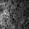
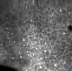
Images are displayed interactively, allowing investigation of dynamic biological processes such as microvascular changes and extravasation. The non-invasive nature of LMI means that tissue sites can be imaged many times. This opens new and unprecedented research opportunities, including monitoring longterm processes.

In-vivo reflectance and fluorescence images

  • Reflectance imaging is based on collecting light scattered from the tissue. Scattering is produced by naturally occurring contrast agents, such as keratin and melanin, or refractive index variations within and between cells. Such features allow the Stratum to be used to examine pigmented lesions and keratinised tissue.
  • For in vivo fluorescence imaging of skin, commonly used contrast agents may be visualised.

Applications with the OptiScan Stratum

  • examination of pigmented or non-pigmented lesions
  • microscopic investigation of treatments
  • evaluation of inflammatory lesions such as psoriasis and eczema

mhtml:file://C:\Documents%20and%20Settings\brian\Desktop\brianwebsite\docs\LiveMicroImagingwithOptiScanStratum.mht!http://www.lawrenz.com/images/stratum_papillary_dermis_thumb.jpg  mhtml:file://C:\Documents%20and%20Settings\brian\Desktop\brianwebsite\docs\LiveMicroImagingwithOptiScanStratum.mht!http://www.lawrenz.com/images/stratum_basale_thumb.jpg  mhtml:file://C:\Documents%20and%20Settings\brian\Desktop\brianwebsite\docs\LiveMicroImagingwithOptiScanStratum.mht!http://www.lawrenz.com/images/stratum_corneum_thumb.jpg  mhtml:file://C:\Documents%20and%20Settings\brian\Desktop\brianwebsite\docs\LiveMicroImagingwithOptiScanStratum.mht!http://www.lawrenz.com/images/stratum_granulosum_thumb.jpg  mhtml:file://C:\Documents%20and%20Settings\brian\Desktop\brianwebsite\docs\LiveMicroImagingwithOptiScanStratum.mht!http://www.lawrenz.com/images/stratum_spinosum_thumb.jpg 

order written information»