Melanosome Movies
To create movies of in vitro melanosomes in motion, I constructed a Matlab GUI that would assist in the removal of the most common artifacts attendant to the imaging of cell cultures. A series of corrected, high resolution, digital images could then be stitched together to form a time-lapse movie. At the time, the ability of the camera system on the microscope to capture and store images limited the temporal capture interval to 3 seconds or longer (much better performance is available today). At the very high spatial resolution of some of the movies, these durations often resulted in the perception of jerky motion due to the appreciable distances the melanosomes traversed between successive shots.
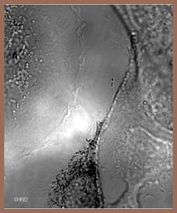
Motivated by the pattern of activity of the melanosomes in these movies, Scott et al. (2002) presented evidence for transfer of melanosomes from melanocytes to keratinocytes through filopodia. Three examples of melanocyte-keratinocyte co-culture movies are available here. Movie1 (6.0 MB) shows a melanocyte dendrite packed with melanosomes and follows the dendrite to a point where, in close-up, it can be viewed in apposition to a keratinocyte. Movie2 (15.9 MB) shows a larger region of a melanocyte in contact with a keratinocyte. Of additional interest in this interaction is the extensive anchoring that holds the two cells in proximity midst all the dynamics.
Movie3 (18.6 MB) contains the original ‘Death Star’ melanocyte segment. Even with the Nomarski imaging, there is only minimal direct visual evidence of the presence of the filopodia. Note the bidirectional movement of the melanosomes as they outline the position of filopodia. This bidirectionality seems to be in contradiction with the reports of unidirectional movement through the filopodia to the keratinocytes by Singh et al. (2008) in their study confirming melanosome transfer. However, much or all of this may well be attributed to experimental differences in the melanocyte-keratinocyte co-cultures. Note also that over the time course of this sequence that the dendrite empties out of melanosomes with the exception of those trapped at the tip by apparent cross-linking. One of the benefits of this display method is that you can go back and create similar high resolution movies of synchronous activity at a different region of interest.
I had initially intended these movies to be a testbed for the use of deconvolution to sharpen high resolution DIC images as the raw images became blurred. However, the discovery of a novel melanosome transport mechanism took precedence and the technology work went back on a crowded shelf of pending projects.