Body Mapping Pilot Images
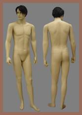
The top level of the of the body mapping representation is a front-back composite of Paolo2, a life-size (1.8 meter) mannequin. The original image was 2612 by 1802 pixels. The next three levels show examples of increasing amounts of magnification. This is one more level than is commonly called for in imaging guidelines.

The next level is a full-front facial image. This is an example of a contextual image that can serve to unambiguously locate a lesion or rash with adequate resolution to identify the condition and a sufficient field of view to record distinguishing anatomical landmarks.
The third level displays a portion of his right eye and lower periorbital skin. This image level commonly captures regions from a few to up to ten centimeters on a side. With cameras that find their way into the exam room commonly providing from 5 to 15 megapixels, this field of view is usually sufficient to convey the necessary diagnostic detail.

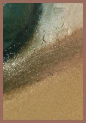
The fourth, and final level is an higher resolution macro shot of the lower eyelid. This image illustrates one reason why Paulo2 has such good looking skin. The surface irregularities mimic the nonplanarity of the stratum corneum and contributes to a more realistic combination of specular and intrinsic reflectance. The field of view is approximately 1.5 by 1.0 millimeters. It will be of interest to determine the degree to which additional diagnostic information can be gleaned from the extra resolution given the presence of motion and focus blur as well as the limitations of the available optics and sensors. The original image was 694 by 478 pixels. A single square pixel covered about 4 micrometers² and was about 4 times the mean wavelength of the captured light across the field of view. Cameras with much higher resolution exist that can be coupled with this lens. Although DIC affords additional imaging capabilities beyond simple transillumination, it is likely that physical limits will soon be reached, if they haven’t been already. Nonetheless, it is expected that although the obtained benefits will vary with the specific diagnostic detail being sought and with the different algorithms being used to process them, performance improvements still have room to increase.
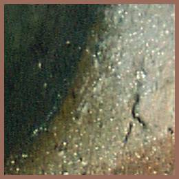

To provide a feeling for the increase in available detail between the whole body shot and a diagnostic close-up image using the same camera, the equivalent representations of a 1 centimeter² region of the mannequin’s eye are presented. A 180 by 180 pixel image is shown next to the average value of its 32,400 pixels. The gray square on the right is the corresponding size of a top level, whole body image pixel when projected onto the same region of the mannequin.